Search This Blog

DEATH AND ITS CAUSES

DEFINITION OF DEATH

Death is the cessation of life in a previously living organism
Medically and scientifically, death is not an event, it is a process. Historically death meant heart and respiration death but heart-lung bypass machine favored new concept ‘brain death’.
There are two aspects of death:
Clinical death
Cellular death

CELLULAR DEATH

means the cessation of respiration (the utilization of oxygen) and the normal metabolic activity in the body tissues and cells.
Cessation of respiration is soon followed by autolysis and decay
Skin and bone will remain metabolically active and thus be ‘alive’ for many hours
The cortical neuron, on the other hand, will die after only 3–7 minutes of complete oxygen deprivation.

SOMATIC DEATH/ CLINICAL DEATH:

The complete and irreversible stoppage of circulation, respiration and brain function.
No legal definition of death.
A person who can not survive upon withdrawal of artificial maintenance is dead.
The individual is irreversibly unconscious and unaware of both the world and his own existence.
The question of death is important in resuscitation and organ transplantation.
Cornea can be removed from the dead body within 6 hours, skin 24 hours, bone 48 hours, kidney and heart obtained soon after circulation has stopped.

Brain death is of 3 types:
  1. Cortical or cerebral death with intact brain-stem, vegetative state.
  2. Brain stem death- where the cerebrum will be intact, though cut off functionally.
  3. Whole brain death (1+2)

VEGETATIVE STATE:

If the cortex alone is damaged, patient passes into coma but the brain stem will function to maintain spontaneous respiration.
Death may occur months or year later.
They are not in need of life sustaining treatment but require nutrition and hydration.
Has stable circulation and shows cycles of eye opening and closing but is unaware of the self and environment.

BRAIN STEM DEATH:

1.The patient must be in deep coma.
2.Treatable causes such as depressant drugs, metabolic or
endocrine disorders (diabetic or myxoedema coma)
or hypothermia must be excluded.
3. The patient must be on mechanical ventilation, no movement and no spontaneous breathing.
4. Cessation of spontaneous cardiac rhythm.
5. No brain stem reflexes- corneal, vestibulo-occular, pupillary, grimacing, gag reflex.
6. Bilateral fixed dilated pupil.


DEATH CERTIFICATION

The format of certifying the cause of death is now defined by the World Health Organization (WHO) and is an international standard that is now used in most countries.


The system divides the cause of death into two parts:
Part I describes the condition(s) that led directly to death;
Part II is for other conditions, not related to those listed in Part I, that have also contributed to death.


The pathologist, forensic medical examiner and senior police officer at the scene of a sudden death.
Mode of death:

According to Bichat, there are 3 modes of death based on death begins in which of the 3 systems: (irrespective of what the remote cause of death may be)

Coma
Syncope
Asphyxia

According to Gordon (1994), the stoppage of vital functions depend upon tissue anoxia. It may be anoxic anoxia (strangulation, choking, drowning, hanging), anaemic anoxia (acute massive hemorrhage, CO), stagnant anoxia (heart failure, shock) and histotoxic anoxia (acute cyanide poisoning).
Anoxic anoxia due to lack of oxygen in the inspired air or mechanical obstruction to respiration is k/a asphyxia.

Coma:
  • State of unarousable unconsciousness.
  • Clinical symptom and not a cause of death.
  • Causes of coma are compression of brain, drugs like opium, cocaine, alcohol; metabolic disorders like uraemia, diabetes.
  • At autopsy, the lungs, brain and the meninges are conjusted, injuries of the brain or the disease may be present.


Syncope:
  • Sudden stoppage of the action of heart, may be fatal.
  • Is due to vaso-vagal attack from reflex para-sympathetic action
  • Blood pressure falls suddenly causing cerebral anemia and rapid unconsciousness, recovery is common.
  • Autopsy of syncope:
  • Heart is contracted and the chambers are empty when d/t anemia,
  • Chambers contain blood when d/t weakness of heart muscles,
  • The lungs, brain and abdominal organs are pale, capillaries conjusted.

Asphyxia:
  • Lack of oxygen in respired air or mechanical obstruction due to which the organs and tissues are deprived of oxygen.
  • Is a mode of death rather than a cause of death.
  • The rule of thumb is : breathing stops within 20 seconds of cardiac arrest and heart stops within 20 minutes of stopping of breathing.

Types of asphyxia:

  • Mechanical- closing of the nose and mouth with hand or mouth, hanging, drowning,
  • Pathological- entry of oxygen is prevented by the disease of URT or lungs eg. bronchitis, acute epiglotitis,
  • Toxic- eg. CO poisoning, opium paralysing respiratory center.
  • Environmental- eg. enclosed places, CO, CO2, high altitude
  • Traumatic- eg. pulmonary embolism,
  • Postural- eg. alcoholic lying down with upper half of the body lower than the remainder
  • Iatrogenic- associated with anaesthesia.

The ‘classical’ features of asphyxia are found where the air passages are obstructed by pressure applied to the neck or to the chest and where there has been a struggle to breathe. The classical features of ‘asphyxia’ are:

1.congestion of the face;
2. oedema of the face;
3.cyanosis (blueness) of the skin of the face;
4.petechial haemorrhages in the skin of the face and the eyes.
5.A fifth feature – increased fluidity of the blood – is now not accepted.


Petechial haemorrhages on the eyelid, conjunctiva in a case of manual strangulation.

Causes of death:

Disease or injury responsible for starting the sequence of events, which are brief or prolonged and which produce death.

Divided into:

Immediate cause- terminal events eg. trauma, peritonitis etc.
Basic cause- pathological process responsible for death at the time of terminal event eg. gunshot wound
Contributory cause- pathological process involved in or complicating but not causing the terminal event.

Manner of death:

Way in which the cause of death was produced.
May be natural or unnatural, violent.
Violence may be of suicidal, homicidal, accidental or undetermined origin.

Mechanism of death:
Congestion is the red appearance of the skin of the face and head, due to the filling of the venous system
Oedema is the swelling of the tissues due to transudation of fluid from the veins caused by the increased venous pressure as a result of obstruction of venous return to the heart.
Cyanosis is the blue colour imparted to the skin by the presence of deoxygenated blood in the congested veins.
Petechial haemorrhages (petechiae) are tiny, pinpoint haemorrhages, most commonly seen in the skin of the head and face and especially in the lax tissues of the eyelids. They are also seen in the conjunctivae and sclera of the eye.

Physiological or biochemical disturbance produced by the cause of death which is incompatible with life eg. shock, sepsis, fibrillation, respiratory paralysis, severe metabolic alkalosis/acidosis.

NEGATIVE AUTOPSY:
When gross and microscopic examination, toxicological analysis and lab investigation fail to reveal the cause of death.
2-5% of all autopsies are neagtive.

SUDDEN DEATH:
When a person not known to have suffering from any dangerous disease, injury or poisoning is found dead, or dies within 24 hours after the onset of terminal illness (WHO)
Some authors call sudden death as occuring instantaneously or within 1 hour of onset of symptoms. Causes are disease of CVS-50%, resp. system-20%, CNS-15%, GI system- 7%, miscellaneous- 5 to 10% eg. DM.

POST-MORTEM CHANGES

IMPORTANCE OF PM FINDINGS

Doctor needs to know the normal progress of decomposition so that he does not misinterpret these normal changes for signs of an unnatural death and,
Secondly, because they can be used in determining how long the individual has been dead.
EARLY CHANGES

Immediate fall in bl. pressure & supply of oxygen ceases.
With loss of neuronal activity, all nervous activity ceases, the reflexes are lost and breathing stops.

In eye,corneal reflex ceases and pupils stop reacting to light.Opacity of cornea d/t drying.

If eyelids open after death, in few hours a film cell debris and mucous form two yellow triangles on scelera on either side of iris, which become brown and then black called ‘tache noir’ within 3-4 hours.

The retinal vessels, viewed with an ophthalmoscope, show the fragmentation of the columns of blood,called ‘trucking’ as it suggests the movement of railway wagons.

The eyes lose their intraocular tension.
The muscles rapidly become flaccid (primary flaccidity), but may respond to touch and other forms of stimulation for some hours after cardiac arrest.

Discharges of the dying motor neurons may stimulate small groups of muscle cells and lead to focal twitching.

The fall in blood pressure and cessation of circulation usually render the skin, conjunctivae and mucous membranes pale. The skin of the face and the lips may remain red or blue in colour in hypoxic/congestive deaths.


Loss of muscle tone in the sphincters may result in voiding of urine; this is such a common finding that no relationship with deaths from epilepsy or asphyxia can be established.

Emission of semen is also found in some deaths; the presence of semen cannot be used as an indicator of sexual activity shortly before death.

Regurgitation is a very common feature of terminal collapse and it is a common complication of resuscitation.


RIGOR MORTIS

occurs within muscle cells as a result of lack of oxygen
In the face of low ATP and high acidity, the actin and myosin fibres bind together and form a gel.
If muscle glycogen levels are low or if the muscle cells are acidic at the time of death as a result of exercise, the process of rigor will develop faster.
Electrocution is also associated with rapidly developing rigor and this may be due to the repeated stimulation of the muscles.
Conversely, in the young, the old or the emaciated, rigor may be extremely hard to detect because of the low muscle bulk.
First detectable in the smaller muscle groups such as those around the eyes and mouth, the jaw and the fingers.

It appears to ‘spread’ down the body from the head to the legs as larger and larger muscle groups are rendered stiff.
Rigor mortis is a chemical process enhanced by heat, so colder the temperature the slower the reactions and vice versa. In a cold body, the onset of rigor will be delayed.
body lying in front of a fire or in a bath of hot water will develop rigor quickly.
In temperate conditions rigor can be first detected (begins)in the face between 1 and 4 hours and in the limbs between 4 and 6 hours after death, and the strength of the rigor increases for the next 6–12 hours. Once established, rigor will remain static until decomposition.
The secondary flaccidity becomes apparent from 24 to 50 hours after death with the onset of putrefaction.
It is best to test for rigor across a joint using very gentle pressure from one or two fingers only.or at eyelids.

A crude but useful aide-memoire is:
Body feels warm and flaccid – dead less than 3 hours.
Body feels warm and stiff – dead 3–8 hours.
Body feels cold and stiff – dead 8–36 hours.
Body feels cold and flaccid – dead more than 36 hours.

CADAVERIC RIGIDITY OR SPASM:
the stiffness of muscles that is said to have its onset immediately at death
Most cases are said to be related to individuals who are at high levels of emotional or physical stress immediately before death
the mechanism for this phenomenon is possibly neurogenic


Cadaveric rigidity – a rare condition. This victim grasped at some ivy as he fell into water.

POST-MORTEM HYPOSTASIS:

The passive settling of red blood cells under the influence of gravity to the blood vessels in lowest areas of the body is important forensically. It produces a pink or bluish colour and it is this colour change that is called post-mortem hypostasis. also called post-mortem lividity and also suggilation.
Hypostasis is not always seen in a body and it may be absent in the young, the old and the clinically anaemic or in those who have died from severe blood loss.
It may be masked by dark skin colours, by jaundice or by some dermatological conditions.

However, hypostasis occurs only where the superficial blood vessels can be distended.
However, compressed areas will remain pale called blanching.
Blanching may also be caused by pressure of clothing or by contact of one area of the body with another
A body left suspended after hanging will develop deep hypostasis of the lower legs and arms, with none visible on the torso, whereas a body that has partially fallen head first out of bed will have the most prominent hypostatic changes of the head and upper chest.the drowning body has different pm lividity.

IMPORTANCE OF LIVIDITY

The cherry pink colour of carbon monoxide poisoning, the dark red or brick red colour associated with cyanide poisoning, and infection by Clostridium perfringens, which is said to result in bronze hypostasis.
The time taken for hypostasis to appear is so variable that it has no significant role in determining the time of death.
Movement of a body will have an effect on hypostasis


Normal distribution of post-mortem hypostasis in a body which lay on its back after death. The white areas are due to pressure upon the ground.


Post-mortem hypostasis on a body found face down on a bed. The linear marks are formed by pressure from creases in the blanket. The pale areas around the mouth and nose are not necessarily signs of suffocation.

COOLING OF THE BODY AFTER DEATH:

Aka algor mortis, is a complex process, not at the same rate through out the body.
Exchange of heat to surrounding occurs only by conduction.
For about half to one hour about death, rectal temperature falls little or not at all. Then, the cooling rate is relatively uniform till 4 degree centigrade.

Postmortem caloricity:
Body temp raised for the 1st 2hrs or so after death, eg. In sunstroke, tetanus, convulsion, septicaemia

Thermometer used to measure rectal temperature is 25 cm long, with a range of 0-50 degree, inserted 8-10cm in rectum for 2mins. The temperature can also be recorded making the hole in the peritoneum and the peritoneal cavity, external auditory meatus, cribiform plate through the nose.
Time since death=normal body temp-rectal temp/rate of temp fall for hours. However, for the formula, we must assume 37 degree rectal temp at the time of death, uniform rate of cooling of whole body.
The rectal temp of an average size naked body reaches the env in about 20hours.

ESTIMATION OF THE TIME OF DEATH:

Of the three historic pathological features that were used to estimate death, only the fall in body temperature has withstood the test of science;
the others – rigor mortis and hypostasis – have now been shown to be utterly unreliable and will not be considered further.

IDENTIFICATION


Identification:

The determination of the individuality of a person based on physical characteristics.
Necessary in living person, recently dead, decomposed body, mutilated body and skeleton.
Necessary in criminal cases like physical assault, murder, rape etc. or in civil cases like marriage, passport, insurance claim, missing person.
At least two identification mark should be noted by the doctor in all ML cases.
The police has to establish the identity of the person. In some cases doctor may be able to supply identification marks of the person, dead body or fragmentary remains.

The corpus delicti: the body of offense, the fact of any criminal offense. The corpus delicti of murder is the fact that the person died from unlawful violence. Clothing with weapon mark, photographs of deceased showing injuries etc. are included in this term.
The main part of corpus delicti is establishment of identification of dead body.
It includes the body of the victim and other facts which are conclusive of dead by foul play, eg bullet or a broken knife found in the body.

Identification Data:
Race determined by
complexion,
eyes (european-blue/gray),
hair (european-fair, brown, straight, wavy; negros-elongated, oval on crosssection with dense pigment; negros have wooly hair; mongolian hair is coarse and dark, usually circular in cross section)
Clothes
Skeleton- the cephalic index=max breadth of skull/ max length*100
dolicocephalic (CI 70-75) aryans, negros,
mesaticephalic (CI 75-80) europeans and chinese
brachycephalic (CI 80-85) mongolian

Sex

In female, barr body present in the buccal mucosa smear, neutrophil content small nuclear attachment of drumstick form (davidson body) in 6%.
Sex determination is difficult in hermaphroditism, concealed sex, advanced decomposition, skeleton.
Males have square orbits, prominent supra-orbital ridges, sub-pubic angle ‘V’ shaped (70-75 degree), pelvic inlet heart shaped, less movable coccyx.

Religion

Hindu males are not circumcised, sacred thread, cast mark on forehead, tuft of hair on the back of head
Hindu females: vermillion in head, silver toe ornaments, nose ring aperture in the left nostril
Muslim females: may have nose ring in septum only, several openings in the ear
Muslim males: circumcised


Age

Can be determined from teeth, ossification of bones, secondary sex characters and general development in case of children.
Temporary and permanent teeth can be seperated. Temporary are smaller, lighter, china-white colored crown, smaller and more divergent roots.
Tooth eruption dates help identify the date
Gustafsons method is used for estimation of age of adults over 21 years. It is based on attrition (wear and tear of occlusal surface upto pulp), paradentosis (regression of gums and periodontal tissues in advancing age or pathology), cementum aposition (secondary cementum slowly and continuously deposited throughout life forming lines appearing as cross striation in the enamel of teeth and used to count to know the age), root resorption, transparency of the root.


Individual bones ossification centre also used to calculate age eg, symphysis pubis, sternum, hyoid bone, skull, sacrum, vertebra. First bone to ossify is clavicle.


General development and stature

Varies at different time of day by 1 &half to 2 cm.
Malnutrition and old age reduces stature.
On an average body lengthens by 2cm after death.
If the body has dismembered, stature may be determined by length of tip of the middle finger to the tip of opposite middle finger, twice the length of one arm with 30cm added for 2clavicles and 4 cm for sternum

Anthropometric measurement

Includes color of hair, eyes, shape of nose
Body marks as moles, scar, tataoos
Body measurement as height, AP diameter of head, the span of outstretched hands
Complexion
External pecularity eg. mole, birth mark, scar, tatoo mark
Age of scar- 5-6 days, reddish or bluish angry scar; by end of 14 days, scar becomes pale; in 2-6 months, the scar becomes white and glistening, tough and may wrinkle
ML importance of scar is mark for identification, age of scar, shape of scar may identify the weapon.

Superimposition is the technique applied to determine whether the skull is that of person in the photograph
Finger print and foot print

Classification of fingerprints:

Loops (67%) may be radial or ulnar
Whorls (25%)
Arch (6-7%)
Composite
Poroscopy is further study of finger prints which studies microscopic pores on the ridges on fingers formed by sweat glands.
Foot prints (podogram) are also used
Lip print (cheiloscopy) also used
Teeth
Personal effect eg. Cloth, jewellery, pocket content
Handwriting (calligraphy is characteristic of the individual, esp if written rapidly
Speech and voice: certain pecularities of speech eg, stammering, stuttering, lisping may be more evident when talking excitedly
Gait
Memory and education (sometimes useful, esp. incase of imposture)

Liver Ultrasound

Scan Planes: - Upper abdominal transverse scan (to demonstrate the left lobe)
- Right subcostal oblique
- high and extended right intercostal scans.
- paramedian upper abdominal longitudinal scans.

Sonographic Anatomy and Normal Findings:
- The liver exhibits a diaphragmatic surface and a visceral surface.
- Both surfaces meet anteroinferiorly at the sharp inferior hepatic border and posterosuperiorly at the fixed part of the diaphragm.
- The liver is divided anatomically into the right and left lobes, the falciform ligament separating the larger right lobe from the smaller left lobe. The quadrate lobe (segment IV) and the caudate lobe (segment I) belong physiologically to the left lobe.


Segmental anatomy of the liver, diaphragmatic surface. A line between the gallbladder and inferior vena cava divides the liver into right (Segment V–VIII) and left physiologic lobes (Segment I–IV)


Segmental anatomy of the liver, visceral surface. Boundaries of the caudate lobe: upper hepatic border, falciform ligament, portal vein, and vena cava. Boundaries of the
quadrate lobe: lower hepatic border,
falciform ligament, gallbladder,
and portal vein

Scanning Protocol
- Transducer: 2.5–5.0 MHz (depending on the abdominal circumference)
- Right subcostal oblique scan: Ask the patient to take a deep breath and hold it.
- Define the dome of the liver with the diaphragm, hepatic veins, portal venous branches (common hepatic duct), the intrahepatic bile ducts, the gallbladder, and the hepatic parenchyma.


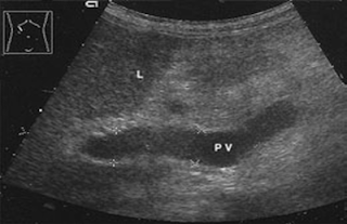
Subcostal oblique scans. Scan through the porta hepatis into the upper part of the liver. PV = right and left branch of the portal vein. V = inferior vena cava, arrow = ligamentum venosum.


Scan directed from the inferior hepatic border (at top of image) to the fixed part (at bottom of image) demonstrates
the quadrate lobe (QL) and caudate lobe (CL) anterior to the vena cava (VC). L = right lobe of liver, PV = portal vein
Schmidt, Ultrasound


Upper abdominal longitudinal scan of the subdiaphragmatic vena cava and the termination of the hepatic veins (arrow). QL = quadrate lobe, PV = portal vein, CL = caudate lobe, VC = inferior vena cava, L = liver


High intercostal scan on the right side demonstrates the costophrenic angle (CA), posterior portions of the diaphragm (DIA) and the entry echo of the lung (L)

Overview of Findings, Classification:
Changes in the liver: Sonographic abnormalities of the liver may consist of diffuse or circumscribed changes in the normal hepatic architecture:
Diffuse changes: These refer to a general alteration of normal liver architecture with regard to size, echogenicity, contours, vasculature, and tubular tracts. Changes in echo texture and contours are particularly significant.
Circumscribed changes: focal alterations in the normal echo texture of the liver. Their detectability depends on the difference in acoustic impedance between the change and normal surrounding liver (anechoic lesions such as cysts are easily recognized). A lesion that is isoechoic to surrounding liver can be distinguished only by the presence of a hypoechoic rim or vascular displacement

Changes in the portal veins: Abnormalities of the portal vein and its tributaries may produce changes identical to those found in the systemic veins.

Diffuse changes in hepatic echogenicity or contours


Circumscribed hepatic changes


1) Fatty Liver


Slight coarsening of the parenchymal echo pattern, increased echogenicity, distal acoustic shadowing, and organ enlargement

2) Chronic Hepatitis


Very slight coarsening of the parenchymal echo pattern and
increased sonodensity with faint acoustic shadowing.
Right subcostal scan

Congestive cirrhosis


Congestive cirrhosis. The liver still has a near normal parenchymal
Echo pattern, but note the curved, bulging inferior border and the tiny
breaks in the capsule (arrows). A = ascites

Very Pronounced changes in echogenicity and contour

1) Sarcoidosis


Sarcoidosis of the liver: coarse parenchymal echo pattern, nonvisualization of
the vessels, and multiple small hypoechoic foci (arrows)

2) Micronodular abscesses or metastases


Micronodular infiltrates in the liver

Anechoic Changes

1) Liver cysts

Solitary Cyst in Liver

Septated Cysts in Liver


Cystic Echinococcosis


Hypoechoic Changes on Liver Ultrasound

1) Focal Sparing in Fatty Infiltration of Liver


Hypoechoic quadrate lobe (segment IV, arrows) adjacent to the gallbladder (GB) in an otherwise fatty liver

2) Regenerative Nodules

Regenerative nodule in severe alcoholic toxic cirrhosis (arrow), confirmed cytologically

3) Hemorrhagic Liver Cyst


4) Liver abscess
Hypoechoic, sharply circumscribed mass with a faintly Echogenic wall


Hyperechoic pyogenic membrane is often present

5) Adenoma

Isoechoic tumor with focal anechoic necrosis / hemorrhage.

Isoechoic Changes in Liver
1) Liver Metastases


The lesions are demarcated from normal liver tissue only by a hypoechoic rim (this accounts for a certain percentage of sonographically occult metastases that are detectable by other modalities).

Echogenic and Hyperechoic Changes


Hemangioma (H) of the liver (L): typical Echogenic, round to oval mass with smooth margins.


Metastasis from colon carcinoma: echogenic round mass with a less echogenic center.


Calcifying metastasis from colorectal carcinoma
Changes in the Portal Venous System

1)Changes in Portal Vein Lumen – Dilatation.

Commonest finding of Portal Hypertension.
But the diagnosis also relies on CDS(colour doppler) and flow patterns across the portal vein lumen
Common causes of Portal Hypertension are: Pre Hepatic – Portal Vein Thrombosis
Intra Hepatic – Cirrhosis
Post Hepatic – Budd Chiari Syndrome

Increased portal vein diameter, indirect signs: - > 11mm intrahepatic, > 13–15mm in the hepatoduodenal ligament
- Caliber variations I 2mm or 50–100 % with respirations
- Detection of hepatic cirrhosis
- Splenomegaly
- Possible ascites
- Wall thickening of the gallbladder and stomach


Incipient portal hypertension. The portal vein (PV) is marginally dilated: 12.9mm intrahepatic

2) Flow Changes and Collaterals:
- Associated with increased portal hypertension.
- Detected on CDS of portal vessels.
Flow velocity is slowed to < 10 cm/s (normal = 15–20 cm/s)
Luminal diameter > 15 mm, does not vary with respirations
Bidirectional, absent or reverse flow in the portal vein or its tributaries


Portal hypertension in liver cirrhosis. CDS: decreased flow velocity
with absence of flow in the portal vein. Flow is in the normal direction (encoded in red), but its velocity is slowed to 9 cm/s. Absence of flow in the portal vein (PV). Additional sign: large-caliber hepatic artery (A), arterial waveform

3) Intraluminal Changes
1)Acute portal or mesenteric vein thrombosis:
- Patient presents with clinical picture of Acute abdomen.
Echogenic filling defect
Vascular dilatation
Absence of color Doppler flow signals

Acute portal vein thrombosis. Mass in the portal vein (VP) is iso echoic to liver tissue (arrows). CDS shows no evidence of flow

2) Chronic Portal Vein Thrombosis


Chronic portal vein thrombosis (PVT) in the setting of a paraneoplastic syndrome. Hepatic metastases: very little increase in luminal diameter, intraluminal echoes in thrombosed portal vein segments. Intrahepatic portal vein is clear

Ultrasound of Gallbladder


Ultrasonic anatomy Located inferior to rt. Lobe of liver
Long axis 6-12 cm , short axis 3-5 cm (4*10cm usually at fasting )
Contracted < 5 cm
Distended > 12 cm when the patient is fasting
Max normal diameter of CBD=4 to 7 mm
Wall Thickness:
Measured in the side in contact with the liver.usually < 3 mm.
From 3-5 mm >>> suspect thick wall
More than 5 mm >>> It is a thick wall gall bladder which is seen in:
Cholecystitis (acute-chronic).
Hepatitis ( viral).
Common Bile Duct is anterior to portal vein.this is the position of porta hapatis

Imaging technique
-overnight fast(6-12hr) to distent GB and remove gas shadow
Short focal length transducer is better as GB is anteriorly located organ.
Supine ,LPO position for imaging
Ganarally dilated bile duct seen in longitudional scan.portal vein ant. To IVC
Contents

Stones:

seen inside, mobile except at the neck they appear white with posterior shadow.
Thick bile.
Called sludge.Change with changing position. The picture occurs in the presence of thick bile in patients on IV fluids for 3-4 days and in inflammation.

Parasite:

Fasciola appears pearl shape.
Move as a whole.

Cancer & polyps:

Polypoidal or heterogeneous mass.


Pathologies

CHOLELITHIASIS-gall stone have high reflictive echo with prominent acoustic posterior shadow
Gravity dependent movement confirms diagnosis
Wall thickening as a hypoechoeic region b/n two echoeic lines,suggest chr.cholecystitis,alcoholism.

Sludge

No acoustic shadow
Low to mild level echoes
Moves very sluggishly
Acute cholecystitis

Associated with cholelithiasis in most cases
Gall stone with focal GB tenderness,usg MURPHY s sign

Pericholecystic edema
Wall edema or wall hypoechoeic
Sometime stone may not be present ,called acalculus cholecystitis eg.burns,old age,after major trauma,and fasting patient

Intrahepatic bile duct

If bile duct are >2mm dia
Second ultrasonic feature of bile duct dilatation is irregularity of bile duct dilatation

Extrahapatic bile duct

Normally 4mm at age 40,5mm at 50
Common site at head of pancrease when double duct sign may be seen
Cbd seen by parasagittal scan

Other facts

Fluid filled colon may appear like GB,wait and watch peristalsis.
In bed ridden ,numerous mass,sludge is not pathological
Polyps donot produce posterior shadow
In obese,difficult to see GB from subcostal approach