Search This Blog

Normal Labour


Case: A 24 year old G2P1, a booked case of your hospital comes to you in labour. She tells you that she has been having labour pain for almost 5 hours now.  On abdominal palpation, you can feel 3 strong uterine contractions every 10 mins. The head is engaged. On PS, cervix is 4cm dilated with effacement. On PV, the head is felt at “0” station. How will you manage the case now? 


What is labour? 

Expulsion of fetus through uterus after the age of viability


What is normal labour ?
  • Vertex presentation
  • Spontaneous onset of labour at term
  • Natural termination through vagina
  • Without any assistance 
  • Without any complications

Onset of labour
  • Fetus – Anterior pituitary and adrenal gland
  • Placenta – Hormones
  • Mother – Uterus, Hormone and Prostaglandins

At term- Increased release of ACTH from the Ant Pituitary
                                    ↓
Increased secretion of cortisol
                 ↓
Increased in oestrogen
                 ↓
Increased oxytocin receptors in the myometrium
                  
Increased prostaglandin synthesis

Prostaglandins
  • Decidua
  • Membranes
  • Placenta
  • Liquor

Diagnosis of onset of labour
  • Painful intermittent uterine contractions 
  • Show
  • Dilatation of cervix
  • Formation of bag of water


Characteristics of uterine contractions

  • Intermittent
  • Regular interval
  • Relaxation in between
  • Followed by retraction


Starts from cornua – descends down
Fundal dominance
Synchronous
Intra amniotic pressure rise to > 20mm Hg
1st stage – 40-50 mm Hg
2nd stage – 100-120 mm Hg 


Characteristics of cervical dilatation

  • Progressive 1cm per hour in primi
  • 1.5 cm per hour in multi
  • Effacement – progressive shortening of cervix due to gradual merging up of upper part of cervix to the lower segment
  • May occur before dilatation of cervix in primi
  • Occurs together with dilatation during labour


Stages of labour

1st stage – Onset of labour to full dilatation of cervix = 10 cm
2nd stage- full dilatation of cervix to delivery of fetus
3rd stage- delivery of fetus to expulsion of placenta

Duration of labour

Total duration 
1st stage -  Primi 8-16 hrs (average 12 hrs)
                -  Multi 4- 8 hrs (average 6 hrs)
2nd stage- Primi 1-2 hrs
                - Multi 1 hr
3rd stage – 5 to 15 min  


Mechanism of labour

  • Engagement
  • Engaging diameter – sub-occipito-bregmatic (9.4 cm)
  • Descent with flexion
  • Internal rotation
  • Crowning
  • Extension
  • Restitution and External rotation
  • Delivery of trunk


Management of 1st stage of labour

1st stage – latent stage and active stage 
General care
Monitor labour

General care

  • Support and care 
  • Mobilization
  • Nutrition
  • Evacuation of bladder
  • Antiseptic and asepsis


Monitor Progress of Labour

Maintain partograph


Components of partograph


  • Mother’s identification
  • Fetal heart rate
  • Color of liquor and moulding
  • Rate of cervical dilatation and descent of head
  • Frequency in 10 min, duration and strength of uterine contraction
  • Vitals = pulse, BP, Temp
  • Oxytocin infusion
  • Drugs and I/V fluids
  • Urine for acetone, albumin and volume


    Management of 2nd stage
    • Position
    • Bearing down during contraction
    • Episiotomy need
    • Support perineum
    • Suction baby’s mouth and nose
    • Keep the baby on mother’s abdomen
    • Dry the baby by wiping
    • Clamp and cut the cord
    • Keep the baby warm
    • Skin to skin contact

    Active Management of 3rd stage
    • Oxytocin 10 units I/M after delivery of fetus
    • Controlled cord traction (CCT) 
    • Uterine massage every 15 min for 2 hrs
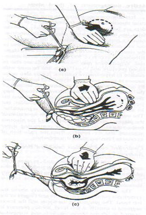


    Mechanism of control of bleeding
    Signs of separation of placenta 
    • Hardening of uterus
    • Gush of blood per vaginum
    • True lengthening of cord
    • Rise of fundal height


    Fetal skull

    Landmark of Fetal skull 

    Occiput:- is the occipital bone/external occipital protuberance.
    Sinciput:- is the forehead region of fetal head.
    Parietal eminences:- are the eminences of parietal bone on either side.
    Mentum:- is the chin.
    Vertical point:- is the center of sagittal suture.
    Frontal point:- is the root of nose.
    Sub occiput:- it is the junction fetal neck and Occiput.
    Sub mentum:- it is the junction between neck and chin.
    Bi parietal:- is the transverse distance between two parietal eminences.
    Bi temporal :- is the distance between two lower end of coronal suture


    SUTURES : SUTURES : 

    Sagittal suture:- 
    This lies in between two parietal bone.
    Coronal suture:- 
    This lies in between the frontal and parietal bone on either side.
    Frontal suture:- 
    This lies in between two frontal bone.
    Lambdoid suture:- 
    It lies in between the parietal and occipital bone on either side.


    CLINICAL IMPORTANCE OF SUTURE

    These suture permit gliding movement of one bone over  other during moulding of the head in the vertex presentation , as a result the diameter of the head get smaller so passage of head through the birth canal become easier.
    Position of fontanelle and sagittal suture can identify attitude and position of vertex.
    From the digital palpation of the sagittal suture during labour, degree of internal rotation and degree of moulding of the head can be noticed.
    In deep transverse arrest, this sagittal suture lies transversely at the level of the ischial spines.


    Area of skull

    A. Vertex:- 
    It is the quadrangular area bounded anteriorly by the bregma and coronal sutures behind by the lambda and the lambdoid sutures and laterally by the line passing through the parietal eminences.
    B. Brow:- 
    It  is an area bounded on one side by the anterior fontanelle and the coronal sutures and on the other side by the root of the nose and supra-orbital ridges of the either side.
    C. Face:- 
    It is an area bounded on one side by the root of the nose and the supra-orbital ridges and on the other by the junction of the floor of mouth with neck.


    Diameter of skull

    The engaging diameter of the fetal skull depends on the degree of the flexion of the presenting part. 

    A. The antero-posterior diameter which may be engaged are:-

    1.Sub-occipito bregmatic:-

    It extends from the nape of the neck to the centre of anterior fontanelle.
    Length:-9.5cm
    Attitude:-complete flexion
    Presentation:-Vertex.

    Clinical importance:- 
    Smallest diameter.


    FETAL SKULL CHANGES IN LABOUR

    Moulding:-It is the changes in shape of the head in vertex presentation during labour while passing through the resistant birth canal.
    Mechanism:-  
    1. Overlapping of cranial bones at the membranous joints due to compression of the engaging diameter of the head.
    2. It is physiological, harmless and disappears within a few hours after birth.


    GRADING

    Grade 0:- the bones lies side by side having an intervening membrane.
    Grade +:- the bone touching but not overlapping
    Grade++:- overlapping but easily separated by pressure.
    Grade+++:- fixed overlapping and cannot be separated.


    CAPUT SUCCEDANEUM

    It is localized area of edema on fetal scalp on vertex presentation due to pressure effect of dilating cervical ring and vaginal introitus.
    Characteristics:-
    It is physiological, present at birth and disappears within 24 hours.
    It is soft, diffuse and pits on pressure.
    No underlying skull bone fracture.


    Cephalhematoma

    It is a collection of blood between periosteum and skull bone which is limited by the periosteal attachments at the suture lines.

    Characteristics:-
    Appears after 12 hours of birth.
    Limited by suture lines.
    Tends to grow larger.
    Disappears within 6-8  weeks.
    It is circumscribed, soft and non pitting.
    May be associated with skull bone fracture.

    Treatment:- No treatment  required. The blood is absorbed and the swelling subside. 


    DIFFERENCES 


    Fetus in-utero

    The lie 

    Relationship between the long axis of the fetus and the long axis of the centralized uterus or maternal spine

    Longitudinal
    long axis of the fetus is aligned to the mother’s
    this is the only NORMAL position

    Transverse
    long axis of the fetus is perpendicular to that of the mother’s

    Oblique
    long axis of the fetus is 0-90 degrees (or 90-180 degrees) to that of the mother’s


    The presentation

    The part of the fetus which occupies the lower pole of the uterus

    Vertex
    head down in the pelvis

    Brow

    Facial

    Breech 
    head is up in the uterine fundus and the buttock in the pelvis

    Shoulder

    Attitude

    Relationship of fetal parts to each other:
    Flexed 
    Deflexed 
    Extended

    Denominator

    Arbitrary bony fixed point on the presenting part

    PRESENTATION

    Vertex
    Brow
    Facial
    Breech
    Shoulder


    ATTITUDE

    Flexed
    Deflexed (vertex)
    Extended (vertex)

    DENOMINATOR

    Occiput
    Frontal eminence
    Mentum
    Sacrum
    Acromion

    Flexed Vertex Presentation 8 Possibilities

    LOL 
    ROL
    LOA
    ROA

    ROP
    LOP 
    OP
    OA

    Leopold’s Maneuver

    Four-part process 
    Aim
    Determine the position of the baby  in utero 
    Determine the expected presentation during labor and delivery 

    Questions to ask yourself when performing the exam:
    • Is the fundal height consistent with the fetal maturity?
    • Is the lie longitudinal, transverse or oblique?
    • Is the presentation cephalic or breech? 
    • If cephalic, is the attitude vertex or facial? 
    • Is the vertex engaged?

    Preparation

    Woman is supine, head slightly elevated and knees slightly flexed
    If the doctor is R handed, stand at the woman’s R side facing her for the first 3 steps, then turn and face her feet for the last step (L handed, left side).


    First Maneuver

    What part is in the fundus?

    Facing the mother, palpate the fundus with both hands
    Assess for shape, size, consistency and mobility

    Fetal head: firm, hard, and round
    Detectable by ballotement

    Buttocks/breech: softer and has bony prominences

    Second Maneuver

    Determine position of the back.

    Still facing the mother, place both palms on the abdomen
    Hold R hand still and with deep but gentle pressure, use L hand to feel for the firm, smooth back 
    Repeat using opposite hands
    Once you’ve located the back, confirm your findings by palpating the fetal extremities on the opposite side  (“lumpy”)

    Third Maneuver

    Determine what part is lying
    above the inlet.

    Gently grasp just above symphisis pubis with the thumb and fingers of the R hand 
    Confirm presenting part (opposite of what’s in the fundus)
    Head will feel firm
    Buttocks will feel softer and irregular
    If it’s not engaged, it may be gently pushed back and forth
    Proceed to the 4th step if it’s not engaged…


    Fourth Maneuver

    Flexed/Deflexed/Extended?

    Turn to face the woman’s feet
    Move fingers of both hands gently down the sides of the abdomen towards the pubis 
    Palpate for the cephalic prominence (vertex)
    Prominence on the same side as the small parts suggests that the head is flexed (optimum)
    Prominence on the same side as the back suggests that the head is extended


    Using a Fetoscope

    Fetal heart Rate (FHR) can be determined by use of:
    Fetoscope 
    specifically designed instruments 
    Clinical stethescope
    Electronic Doppler


    Doppler Method

    Employs a continuous ultrasound
    Can detect the fetal heart at 10-12 weeks’ gestation

    Amplifiers allow both the practitioner and parents to hear


    Fetoscope

    Can pick up the fetal heart rate at 17-19 weeks’ gestation


    Fetal heart tones are best heard over the baby’s back
    Used in conjunction with Leopold’s maneuver
    Auscultation may be difficult if
    Mother is overweight
    Placenta is in the front of the uterus

    *Always easier in later stages of the pregnancy


    Where will you hear the FHTs?


    Fetal Physiology


    Consists of :                            
    Fetal blood
    Pulmonary system
    Immune system
    Skin 
    Nervous system
    Gastro-intestinal  system
    Urinary system
    Endocrine glands
    Cardiovascular

    Fetal blood

    Haematopoesis:
    1st fetal blood cells  : Day 14 embryo : in yolk sac

    Next, major site is liver: 6th wk embryo  : (hence, enlarged liver) & spleen (lesser extent)
    Finally, bone marrow starts to produce RBC:16 wks

    By 26 wks :  bone marrow is the predominant source of RBC

    As the fetus grows, volume of blood in fetoplacental circulation increases.
           At term, 125 ml/kg fetal wt.
    Hb in the fetal blood also rises.
           At term, Hb  18 g/dl. Why ? Enhances    the transfer of O2 across placenta.
    Fetal RBC has shorter lifespan.
           At term, 90 days.


    Hb in fetus:
    Mostly fetal haemoglobin (HbF):    Î±2 γ2  instead of adult Hb A (α2 β2 ) and Hb A2 (α2 δ2 )
       
         10 – 28 wks    90 % of fetal Hb is HbF
         28 – 34 wks    HbF: HbA= 80:20
         6 mths age     HbF = 1 %
    HbF vs HbA
         HbF has greater O2 binding capacity and it is resistant to denaturation by acid/alkali.


    Fetal Haemoglobin
    HbF  has higher affinity for O2 than adult in vivo due to a lower sensitivity to DPG (2,3 diphosphoglyceric acid)

    Partial pressure of O2 in fetal circulation is low (92.7KPa) but this is compensated for by high Hb concentration and greater oxygen affinity


    CVS

    Differences (fetal vs adult circulation):

    Major portion of Rt ventricular output bypasses the lungs coz fetal blood does not need to enter pulmonary vasculature
    Oxygenation occurs in the placenta
    Right & Left ventricles work in parallel ,rather than in series
    Heart, brain & upper body receive blood from left ventricle ; placenta & lower body receive blood from both right & left ventricles

    Presence of shunts:
             1. Ductus Venosus
             2. Foramen ovale
             3. ductus arteriosus

    Fetal Circulation

    Fetal blood pathway


    Placenta  ----   umbilical vein 
    ----
          ductus      venosus  
    ----
            IVC 
    ----
          right atrium  
    ----
          foramen ovale  
    ----
          left atrium  
    ----
           left      ventricle   
    ----
           ascending aorta
    •    coronary/cerebral arteries       SVC      right atrium       right ventricle       pulmonary artery        ductus arteriosus       descending aorta        umbilical artery      placenta


    After birth, constriction/collapse of:
     umbilical arteries: instantaneous functional closure / actual obliteration takes 2-3 mths. Distal part – form lateral umbilical ligament. Proximal part – remain as superior vesical arteries

    Umbilical veins: closure a little later than arteries. Forms ligamentum teres.
    ductus arteriosus: functional closure soon after pulmonary circn is established. Anatomical obliteration takes 1-3 mths. Forms ligamentum arteriosum.

    foramen ovale: functional closure soon after birth, anatomical closure in 1 yr.

    ductus venosum: forms ligamentum venosum


    Incidence of closure of fetal channels
    Fetal circulatory response to hypoxia
    • Heart rate falls
    • Resistance in the umbilical artery increases
    • Resistance in the middle cerebral artery decreases thus protecting flow to the fetal brain
    • Blood flow increased to heart and adrenals
    • Blood flow reduced to kidneys producing oligohydramnios (reduced volume of amniotic fluid)


    Control of Fetal Heart Rate
    Control of FHR is complex.

    It is subject to modulating influences such as catecholamines and baroreceptors.
    These influences generally act on FHR via the autonomic nervous system.

    Parasympathetic tone dominates


    Fetal Heart rates (FHR):
    -20 weeks 155/minute
    -30 weeks 144minute
    -Term 140/min


    FHR decreases in response to hypoxia


    Fetal  Lung and Breathing


    Development consist of

    Pseudoglandular stage: 5-16 w
    Canalicular stage: 16-25 w
    Terminal sac stage: type II cells begin to produce surfactant 
    At birth only 15% of adult alveoli are present in lungs       continue to grow up to 8 years


    20 wks: full differentiation of capillary & canalicular elements of fetal lung

    24 wks: alveoli develop, surfactant appear.

       Lung alveoli lined by surfactant (a group of phospholipids). 
       Prevents collapse of small alveoli during expiration by lowering surface tension
       Synthesized by type 2 alveolar cells ( 10 % of lung parenchyma)
       Lecithin ( phosphatidyl choline): main (80%)

    Increased lecithin production :
    Cortisol
    Growth restriction
    Prolonged rupture of membranes

    Delayed lecithin formation in:
    diabetes 

    Phosphatidyl glycerol is more predictive of RDS esp in diabetic fetus.


    Surfactant:

    Composition: 90% lipids, 10% proteins
    Phosphatidylcholine (lecithins):
                      DPPC: active component(50%)
                      PG: second(8-15%)
    At birth,with first breath, an air to tissue interface is produced in the alveolus. Surfactant spreads to line alveolus to prevent collapse during expiration

    Glucocorticoids and Surfactant :

    Accelerate type 2 alveolar cell development

    Accelerate surfactant synthesis

    Accelerate lung structural maturation

    Glucocorticoids play essential role in maturation of many fetal organs but excess amounts reduce fetal growth

    Fetal breathing: 

    Numerous but intermittent fetal breathing
       movements occur in utero especially during 
       sleep ( lung maturation).
    Immune system

    8 wks:  lymphocytes appear

    Mid 2nd trimester: all phagocytic cells, T & B cells, complement

     Fetus consists almost totally of maternal IgG (transferred across placenta).
               16 wks: Maternal IgG transfer begins & increases
                Last 4 wks: bulk of IgG acquired.
                Hence, preterm :  less IgG
                Newborns produce IgG slowly & adult values are   reached only after 3 yrs.

    IgM & IgA: very little is produced by fetus.
               not transferred across placenta.
              

    General immunological defences:

    1.Amniotic fluid (lysosomes, IgG)

    2.Placenta (lymphoid cells, phagocytes, barrier)

    3.Liver ( granulocytes)


    Skin

    16 wks: lanugo appears but disappears near term

    1 month gestation till birth: skin thickness progressively increases

    20 wks:  stratum corneum  distinct

    Last wks: skin covered by vernix caseosa
                       desquamated skin cells
                       rich in cholesterol and glycogen

    Preterm babies
    • No vernix & thin skin ie, larger insensible loss

    • Deficient brown fat

    • Delayed development of sweat glands

    Alimentary system & energy stores
    • 10 wks: Swallowing reflex develops & gradually matures. Continuously and increasingly swallows amniotic fluid

    • 2nd trimester:  Peristalsis in intestines

    • Term : Large bowel is filled with meconium
        
    • But defaecation in utero (meconium in amniotic fluid) unusual unless fetal anoxia

    Intestinal villi well developed by 19weeks


    • Gut development important for amniotic fluid
        homeostasis; (fetus swallows amniotic fluid
        from 12 weeks and amniotic fluid contains
        hormones and growth factors that stimulate
        gut development)

    • Gastrin, motilin and somatostatin regulate
        growth and development      present in gut by 13
        weeks         matures by 24 weeks

    • Digestive enzymes eg disaccharidases
        present by 9-10 weeks maturity at term

    Meconium

    Lanugo, vernix, scalp hair, epithelial cells from skin, mucus, exfoliated intestinal epithelium, intestinal juices
        
    Liver:

    Reduced capacity for conjugation of bilirubin esp. in preterm

    Preterm infants:

    Virtually no fat, so severely reduced ability to withstand starvation
    Incompletely developed alimentary system
    Poor and unsustained sucking ability
    Uncoordinated swallowing mechanism
    Delayed gastric emptying
    Poor absorption of carbohydrates/fat/other nutrients

    Kidney & urinary tract
    • Urine usu found in UB of even small fetuses

    • Fetal urine  gives rise to much of amniotic fluid. 
       (protein-free & sugar-free hypotonic ultrafiltrate of fetal plasma)
           12 wks:  urine production starts
           32 wks:  12 ml per hour
           40 wks:   38 ml per hour 

    • 36 wks: nephrogenesis complete
       But maturation of excretory & concentrating ability of fetal kidneys is gradual

       Preterm: immature, so abnormal

    Amniotic fluid

    Source:
    • Early pregnancy: ultrafiltrate of maternal plasma
    • 10 wks: transudate of fetal serum via skin & umbilical cord
    •  After 20 wks: mainly fetal urine ( as cornification of fetal skin, impermeable to water).
        Pulmonary fluid and fluid filtering through  placenta contribute little.

    • AF volume:

            Varies at each week of gestation. 
            Volume increases with increasing    gestation.
            10 wks: 30 ml
            20 wks: 300 ml
            30 wks: 600 ml
            38 wks: 1000 ml
            40 wks:  800 ml
            42 wks:  350 ml

    Functions of AF:
    • Cushions fetus: Protect fetus from mechanical injury
    • Permits movements of fetus while preventing limb contracture
    • Prevents adhesions between fetus & amnion
    • Maintains temperature
    • Minimal nutritive function
    • Permit fetal lung development--  2 way movements of fluid into fetal bronchioles
    •    Absence of AF in 2nd trimester:  pulmonary hypoplasia
    • Promotes growth & differentiation of GI tract 

    • Renal agenesis, cystic kidney, IUGR ----oligohydramnios----reduced contribution of fluid into amniotic sac ----- major alterations in AF volume.

    • Anencephaly, oesophageal atresia, duodenal atresia ----- polyhydramnios----- reduced removal of fluid

    Fetal behaviour
    • 18 wk: first fetal movements (quickening) perceived in primipara
        Several wks earlier in multipara
    • Formal counting of fetal movements (screening)

    Reduced or Absent in 

    • Chronic hypoxia & growth failure
    • fetal death

    • Spinal cord extends along entire length of vertebral column in embryo, but after that, it grows more slowly.
        By 24 wks:  spinal cord extends to S1
        At birth:  L3
        In adults:  L1
    • Myelination of spinal cord:  starts mid-preg
    • 10 wks: swallowing
    • 14-16 wks: respiration evident
    • After 24 wks: ability to suck
    • 24-25 wks: can hear some sounds in utero
    • 28 wks: sensitive to light

    Fetal endocrinology
    • 10 wks: GH, ACTH, PRL , TSH produced by fetal pituitary
    • 1st trimester: Vasopressin, oxytocin from posterior pituitary
    • Fetal adrenal: hypertrophy of reticular zone (site of synthesis of DHEA-dehydroepiandrosterone). This zone is absent in anencephaly. Adrenal medulla produces catecholamines.
    • 11 wks: Fetal thyroid produces small amount thyroxine
    • Fetal ovaries remain inactive


    • Fetal testicles mediate development of male reproductive structures


    • 12 wks: fetal pancreas secrete insulin


    PLACENTA AND FETAL MEMBRANES


    PLACENTA
    • Development
    • Gross anatomy
    • Structures
    • Functions
    • Abnormalities

    DEVELOPMENT

    1.Chorionic frondosum- fetal part the major component
    2.Decidua basalis- maternal part the minor component

    Begins at 6th weeks of gestation
    Ends at 12th weeks of gestation

    Anatomy
    • Normally situated at or near fundus of uterus
    • Shape- discoid
    • Size- 15*20 cms, 2.5cm in thickness in centre
    • 500gms in weight
    • Two surfaces

    1.Fetal- smooth, amniotic membrane, cord insertion and branching vessels.
    2.Maternal- irregular,15-20 cotyledons,

    Fetal surface

    Maternal surface


    Structure


    Functions
    1. Nutrition
    2. Respiration
    3. Excretion
    4. Barrier
    5. Hormonal- CRH, GnRH, TRH, GHRH, ACTH, hCG, hPL, hCT
    6. Immunological- placental hormones, proteins(SP1), early pregnancy factor(EPF), steroids and hCG

    Abnormal placenta

    One or more smaller lobes of placenta placed at varying distances from placental margin.

    Smaller chorionic plate than basal plate.


    Cord Abnormalities

    The cord is attached to the membranes.

    The cord is attached to the margin of the placenta.


    FETAL MEMBRANES
    • CONSISTS OF 2 LAYERS

    CHORION

    AMNION

    FUNCTIONS OF MEMBRANES

    1.PRODUCES AMNIOTIC FLUID

    2.PREVENTS FETUS FROM INFECTIONS

    3.HELPS IN CERVICAL DILATION IN LABOR

    4.PRODUCES PGE2, PGF2alpha